| 中文名稱 | 磷酸化腺苷單磷酸活化蛋白激酶α2抗體 |
| 別 名 | PRKAA1(phospho T172); AMPK alpha 1 + AMPK alpha 2 (phospho T172); phospho-AMPK alpha-1 (Thr183); 5'-AMP-activated protein kinase catalytic subunit alpha-2; AAPK2_HUMAN; AAPK1_HUMAN; ACACA kinase; Acetyl-CoA carboxylase kinase; AMPK alpha 2 chain; AMPK subunit alpha-2; AMPK2; AMPK 2; AMPKa2; AMPK a2; AMPK-a2 AMPKalpha2; HMGCR kinase; Hydroxymethylglutaryl-CoA reductase kinase; PRKAA; PRKAA2; Protein kinase AMP activated alpha 2 catalytic subunit; Protein kinase AMP activated catalytic subunit alpha 2. |
| 產(chǎn)品類型 | 磷酸化抗體 |
| 研究領(lǐng)域 | 免疫學(xué) 神經(jīng)生物學(xué) 信號轉(zhuǎn)導(dǎo) 轉(zhuǎn)錄調(diào)節(jié)因子 激酶和磷酸酶 糖尿病 Alzheimer's |
| 抗體來源 | Rabbit |
| 克隆類型 | Polyclonal |
| 交叉反應(yīng) | Human, Mouse, Rat, Sheep, (predicted: Chicken, Dog, Pig, Cow, Horse, ) |
| 產(chǎn)品應(yīng)用 | WB=1:500-2000 ELISA=1:500-1000 IHC-P=1:100-500 IHC-F=1:100-500 Flow-Cyt=0.2μg /test IF=1:100-500 (石蠟切片需做抗原修復(fù)) not yet tested in other applications. optimal dilutions/concentrations should be determined by the end user. |
| 分 子 量 | 64kDa |
| 細胞定位 | 細胞核 細胞漿 |
| 性 狀 | Liquid |
| 濃 度 | 1mg/ml |
| 免 疫 原 | KLH conjugated Synthesised phosphopeptide derived from human AMPK alpha 2 around the phosphorylation site of Thr172:LR(p-T)SC |
| 亞 型 | IgG |
| 純化方法 | affinity purified by Protein A |
| 儲 存 液 | 0.01M TBS(pH7.4) with 1% BSA, 0.03% Proclin300 and 50% Glycerol. |
| 保存條件 | Shipped at 4℃. Store at -20 °C for one year. Avoid repeated freeze/thaw cycles. |
| PubMed | PubMed |
| 產(chǎn)品介紹 | The protein encoded by this gene is a catalytic subunit of the AMP-activated protein kinase (AMPK). AMPK is a heterotrimer consisting of an alpha catalytic subunit, and non-catalytic beta and gamma subunits. AMPK is an important energy-sensing enzyme that monitors cellular energy status. In response to cellular metabolic stresses, AMPK is activated, and thus phosphorylates and inactivates acetyl-CoA carboxylase (ACC) and beta-hydroxy beta-methylglutaryl-CoA reductase (HMGCR), key enzymes involved in regulating de novo biosynthesis of fatty acid and cholesterol. Studies of the mouse counterpart suggest that this catalytic subunit may control whole-body insulin sensitivity and is necessary for maintaining myocardial energy homeostasis during ischemia. [provided by RefSeq, Jul 2008]The protein encoded by this gene is a catalytic subunit of the AMP-activated protein kinase (AMPK). AMPK is a heterotrimer consisting of an alpha catalytic subunit, and non-catalytic beta and gamma subunits. AMPK is an important energy-sensing enzyme that monitors cellular energy status. In response to cellular metabolic stresses, AMPK is activated, and thus phosphorylates and inactivates acetyl-CoA carboxylase (ACC) and beta-hydroxy beta-methylglutaryl-CoA reductase (HMGCR), key enzymes involved in regulating de novo biosynthesis of fatty acid and cholesterol. Studies of the mouse counterpart suggest that this catalytic subunit may control whole-body insulin sensitivity and is necessary for maintaining myocardial energy homeostasis during ischemia. [provided by RefSeq, Jul 2008] Function: Catalytic subunit of AMP-activated protein kinase (AMPK), an energy sensor protein kinase that plays a key role in regulating cellular energy metabolism. In response to reduction of intracellular ATP levels, AMPK activates energy-producing pathways and inhibits energy-consuming processes: inhibits protein, carbohydrate and lipid biosynthesis, as well as cell growth and proliferation. AMPK acts via direct phosphorylation of metabolic enzymes, and by longer-term effects via phosphorylation of transcription regulators. Also acts as a regulator of cellular polarity by remodeling the actin cytoskeleton; probably by indirectly activating myosin. Regulates lipid synthesis by phosphorylating and inactivating lipid metabolic enzymes such as ACACA, ACACB, GYS1, HMGCR and LIPE; regulates fatty acid and cholesterol synthesis by phosphorylating acetyl-CoA carboxylase (ACACA and ACACB) and hormone-sensitive lipase (LIPE) enzymes, respectively. Regulates insulin-signaling and glycolysis by phosphorylating IRS1, PFKFB2 and PFKFB3. AMPK stimulates glucose uptake in muscle by increasing the translocation of the glucose transporter SLC2A4/GLUT4 to the plasma membrane, possibly by mediating phosphorylation of TBC1D4/AS160. Regulates transcription and chromatin structure by phosphorylating transcription regulators involved in energy metabolism such as CRTC2/TORC2, FOXO3, histone H2B, HDAC5, MEF2C, MLXIPL/ChREBP, EP300, HNF4A, p53/TP53, SREBF1, SREBF2 and PPARGC1A. Acts as a key regulator of glucose homeostasis in liver by phosphorylating CRTC2/TORC2, leading to CRTC2/TORC2 sequestration in the cytoplasm. In response to stress, phosphorylates 'Ser-36' of histone H2B (H2BS36ph), leading to promote transcription. Acts as a key regulator of cell growth and proliferation by phosphorylating TSC2, RPTOR and ATG1: in response to nutrient limitation, negatively regulates the mTORC1 complex by phosphorylating RPTOR component of the mTORC1 complex and by phosphorylating and activating TSC2. In response to nutrient limitation, promotes autophagy by phosphorylating and activating ULK1. AMPK also acts as a regulator of circadian rhythm by mediating phosphorylation of CRY1, leading to destabilize it. May regulate the Wnt signaling pathway by phosphorylating CTNNB1, leading to stabilize it. Also phosphorylates CFTR, EEF2K, KLC1, NOS3 and SLC12A1. Subunit: AMPK is a heterotrimer of an alpha catalytic subunit (PRKAA1 or PRKAA2), a beta (PRKAB1 or PRKAB2) and a gamma non-catalytic subunits (PRKAG1, PRKAG2 or PRKAG3). Interacts with FNIP1 and FNIP2. Subcellular Location: Cytoplasm. Nucleus. Note=In response to stress, recruited by p53/TP53 to specific promoters. Post-translational modifications: Ubiquitinated. Phosphorylated at Thr-172 by STK11/LKB1 in complex with STE20-related adapter-alpha (STRADA) pseudo kinase and CAB39. Also phosphorylated at Thr-172 by CAMKK2; triggered by a rise in intracellular calcium ions, without detectable changes in the AMP/ATP ratio. CAMKK1 can also phosphorylate Thr-172, but at much lower lvel. Dephosphorylated by protein phosphatase 2A and 2C (PP2A and PP2C). Phosphorylated by ULK1; leading to negatively regulate AMPK activity and suggesting the existence of a regulatory feedback loop between ULK1 and AMPK. Similarity: Belongs to the protein kinase superfamily. CAMK Ser/Thr protein kinase family. SNF1 subfamily. Contains 1 protein kinase domain. SWISS: P54646 Gene ID: 5563 Database links: Entrez Gene: 5563 Human Entrez Gene: 108079 Mouse Entrez Gene: 78975 Rat Omim: 600497 Human SwissProt: P54646 Human SwissProt: Q8BRK8 Mouse SwissProt: Q09137 Rat Unigene: 437039 Human Unigene: 48638 Mouse Unigene: 64583 Rat Important Note: This product as supplied is intended for research use only, not for use in human, therapeutic or diagnostic applications. |
| 產(chǎn)品圖片 | 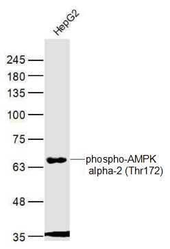 Sample: Sample:HepG2(Human) Cell Lysate at 40 ug Primary: Anti-phospho-AMPK alpha-2 (Thr172) (bs-4002R) at 1/300 dilution Secondary: IRDye800CW Goat Anti-Rabbit IgG at 1/20000 dilution Predicted band size: 63 kD Observed band size: 67 kD 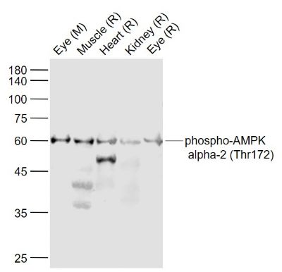 Sample: Sample:Lane 1: Eye (Mouse) Lysate at 40 ug Lane 2: Muscle (Rat) Lysate at 40 ug Lane 3: Heart (Rat) Lysate at 40 ug Lane 4: Kidney (Rat) Lysate at 40 ug Lane 5: Eye (Rat) Lysate at 40 ug Primary: Anti-phospho-AMPK alpha-2 (Thr172) (bs-4002R) at 1/1000 dilution Secondary: IRDye800CW Goat Anti-Rabbit IgG at 1/20000 dilution Predicted band size: 62 kD Observed band size: 60 kD 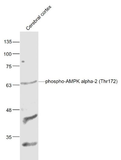 Sample: Sample:Cerebral cortex(Mouse) Lysate at 40 ug Primary: Anti-phospho-AMPK alpha-2 (Thr172) (bs-4002R) at 1/1000 dilution Secondary: IRDye800CW Goat Anti-Rabbit IgG at 1/20000 dilution Predicted band size: 64 kD Observed band size: 64 kD 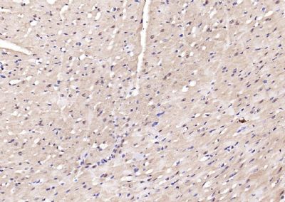 Paraformaldehyde-fixed, paraffin embedded (rat heart); Antigen retrieval by boiling in sodium citrate buffer (pH6.0) for 15min; Block endogenous peroxidase by 3% hydrogen peroxide for 20 minutes; Blocking buffer (normal goat serum) at 37°C for 30min; Antibody incubation with (phospho-AMPK alpha-2 (Thr172)) Polyclonal Antibody, Unconjugated (bs-4002R) at 1:200 overnight at 4°C, followed by operating according to SP Kit(Rabbit) (sp-0023) instructionsand DAB staining. Paraformaldehyde-fixed, paraffin embedded (rat heart); Antigen retrieval by boiling in sodium citrate buffer (pH6.0) for 15min; Block endogenous peroxidase by 3% hydrogen peroxide for 20 minutes; Blocking buffer (normal goat serum) at 37°C for 30min; Antibody incubation with (phospho-AMPK alpha-2 (Thr172)) Polyclonal Antibody, Unconjugated (bs-4002R) at 1:200 overnight at 4°C, followed by operating according to SP Kit(Rabbit) (sp-0023) instructionsand DAB staining.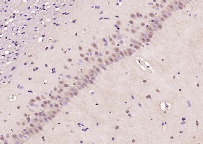 Paraformaldehyde-fixed, paraffin embedded (rat brain); Antigen retrieval by boiling in sodium citrate buffer (pH6.0) for 15min; Block endogenous peroxidase by 3% hydrogen peroxide for 20 minutes; Blocking buffer (normal goat serum) at 37°C for 30min; Antibody incubation with (phospho-AMPK alpha-2 (Thr172)) Polyclonal Antibody, Unconjugated (bs-4002R) at 1:200 overnight at 4°C, followed by operating according to SP Kit(Rabbit) (sp-0023) instructionsand DAB staining. Paraformaldehyde-fixed, paraffin embedded (rat brain); Antigen retrieval by boiling in sodium citrate buffer (pH6.0) for 15min; Block endogenous peroxidase by 3% hydrogen peroxide for 20 minutes; Blocking buffer (normal goat serum) at 37°C for 30min; Antibody incubation with (phospho-AMPK alpha-2 (Thr172)) Polyclonal Antibody, Unconjugated (bs-4002R) at 1:200 overnight at 4°C, followed by operating according to SP Kit(Rabbit) (sp-0023) instructionsand DAB staining.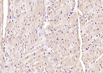 Paraformaldehyde-fixed, paraffin embedded (mouse heart); Antigen retrieval by boiling in sodium citrate buffer (pH6.0) for 15min; Block endogenous peroxidase by 3% hydrogen peroxide for 20 minutes; Blocking buffer (normal goat serum) at 37°C for 30min; Antibody incubation with (phospho-AMPK alpha-2 (Thr172)) Polyclonal Antibody, Unconjugated (bs-4002R) at 1:200 overnight at 4°C, followed by operating according to SP Kit(Rabbit) (sp-0023) instructionsand DAB staining. Paraformaldehyde-fixed, paraffin embedded (mouse heart); Antigen retrieval by boiling in sodium citrate buffer (pH6.0) for 15min; Block endogenous peroxidase by 3% hydrogen peroxide for 20 minutes; Blocking buffer (normal goat serum) at 37°C for 30min; Antibody incubation with (phospho-AMPK alpha-2 (Thr172)) Polyclonal Antibody, Unconjugated (bs-4002R) at 1:200 overnight at 4°C, followed by operating according to SP Kit(Rabbit) (sp-0023) instructionsand DAB staining.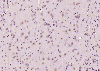 Paraformaldehyde-fixed, paraffin embedded (mouse brain); Antigen retrieval by boiling in sodium citrate buffer (pH6.0) for 15min; Block endogenous peroxidase by 3% hydrogen peroxide for 20 minutes; Blocking buffer (normal goat serum) at 37°C for 30min; Antibody incubation with (phospho-AMPK alpha-2 (Thr172)) Polyclonal Antibody, Unconjugated (bs-4002R) at 1:200 overnight at 4°C, followed by operating according to SP Kit(Rabbit) (sp-0023) instructionsand DAB staining. Paraformaldehyde-fixed, paraffin embedded (mouse brain); Antigen retrieval by boiling in sodium citrate buffer (pH6.0) for 15min; Block endogenous peroxidase by 3% hydrogen peroxide for 20 minutes; Blocking buffer (normal goat serum) at 37°C for 30min; Antibody incubation with (phospho-AMPK alpha-2 (Thr172)) Polyclonal Antibody, Unconjugated (bs-4002R) at 1:200 overnight at 4°C, followed by operating according to SP Kit(Rabbit) (sp-0023) instructionsand DAB staining.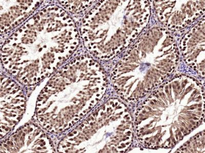 Paraformaldehyde-fixed, paraffin embedded (Rat testis); Antigen retrieval by boiling in sodium citrate buffer (pH6.0) for 15min; Block endogenous peroxidase by 3% hydrogen peroxide for 20 minutes; Blocking buffer (normal goat serum) at 37°C for 30min; Antibody incubation with (phospho-AMPK alpha-2 (Thr172)) Polyclonal Antibody, Unconjugated (bs-4002R) at 1:400 overnight at 4°C, followed by operating according to SP Kit(Rabbit) (sp-0023) instructionsand DAB staining. Paraformaldehyde-fixed, paraffin embedded (Rat testis); Antigen retrieval by boiling in sodium citrate buffer (pH6.0) for 15min; Block endogenous peroxidase by 3% hydrogen peroxide for 20 minutes; Blocking buffer (normal goat serum) at 37°C for 30min; Antibody incubation with (phospho-AMPK alpha-2 (Thr172)) Polyclonal Antibody, Unconjugated (bs-4002R) at 1:400 overnight at 4°C, followed by operating according to SP Kit(Rabbit) (sp-0023) instructionsand DAB staining.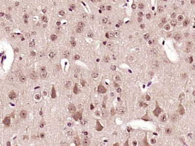 Paraformaldehyde-fixed, paraffin embedded (Mouse brain); Antigen retrieval by boiling in sodium citrate buffer (pH6.0) for 15min; Block endogenous peroxidase by 3% hydrogen peroxide for 20 minutes; Blocking buffer (normal goat serum) at 37°C for 30min; Antibody incubation with (phospho-AMPK alpha-2 (Thr172)) Polyclonal Antibody, Unconjugated (bs-4002R) at 1:400 overnight at 4°C, followed by a conjugated secondary antibody (sp-0023) for 20 minutes and DAB staining. Paraformaldehyde-fixed, paraffin embedded (Mouse brain); Antigen retrieval by boiling in sodium citrate buffer (pH6.0) for 15min; Block endogenous peroxidase by 3% hydrogen peroxide for 20 minutes; Blocking buffer (normal goat serum) at 37°C for 30min; Antibody incubation with (phospho-AMPK alpha-2 (Thr172)) Polyclonal Antibody, Unconjugated (bs-4002R) at 1:400 overnight at 4°C, followed by a conjugated secondary antibody (sp-0023) for 20 minutes and DAB staining.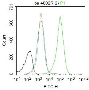 Blank control:Rat spleen. Blank control:Rat spleen.Primary Antibody (green line): Rabbit Anti-AMPK alpha-1 antibody (bs-4002R) Dilution: 2μg /10^6 cells; Isotype Control Antibody (orange line): Rabbit IgG . Secondary Antibody : Goat anti-rabbit IgG-AF488 Dilution: 1μg /test. Protocol The cells were fixed with 4% PFA (10min at room temperature)and then permeabilized with 90% ice-cold methanol for 20 min at room temperature. The cells were then incubated in 5%BSA to block non-specific protein-protein interactions for 30 min at room temperature .Cells stained with Primary Antibody for 30 min at room temperature. The secondary antibody used for 40 min at room temperature. Acquisition of 20,000 events was performed. 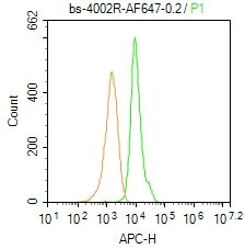 Blank control: Mouse spleen. Blank control: Mouse spleen.Primary Antibody (green line): Rabbit Anti-phospho-AMPK alpha-2 (Thr172)/AF647 Conjugated antibody (bs-4002R-AF647) Dilution: 0.2μg /10^6 cells; Isotype Control Antibody (orange line): Rabbit IgG-AF647 . Protocol The cells were fixed with 4% PFA (10min at room temperature)and then permeabilized with 90% ice-cold methanol for 20 min at-20℃. The cells were then incubated in 5% BSA to block non-specific protein-protein interactions for 30 min at room temperature. The cells were stained with Primary Antibody for 30 min at room temperature. Acquisition of 20,000 events was performed. 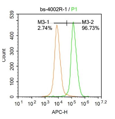 Blank control:Mouse spleen. Blank control:Mouse spleen.Primary Antibody (green line): Rabbit Anti-phospho-AMPK alpha-2 (Thr172) antibody (bs-4002R) Dilution: 1μg /10^6 cells; Isotype Control Antibody (orange line): Rabbit IgG . Secondary Antibody: Goat anti-rabbit IgG-AF647 Dilution: 1μg /test. Protocol The cells were fixed with 4% PFA (10min at room temperature)and then permeabilized with 90% ice-cold methanol for 20 min at-20℃. The cells were then incubated in 5%BSA to block non-specific protein-protein interactions for 30 min at at room temperature .Cells stained with Primary Antibody for 30 min at room temperature. The secondary antibody used for 40 min at room temperature. Acquisition of 20,000 events was performed. 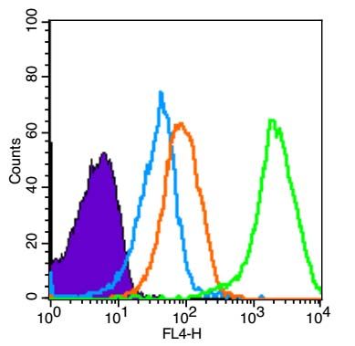 Blank control (Black line): Mouse spleen(Black). Blank control (Black line): Mouse spleen(Black).Primary Antibody (green line): Rabbit Anti-phospho-AMPK alpha-1 (Thr172) antibody (bs-4002R) Dilution: 3μg /10^6 cells; Isotype Control Antibody (orange line): Rabbit IgG . Secondary Antibody (white blue line): Goat anti-rabbit IgG-AF647 Dilution: 1μg /test. Protocol The cells were fixed with 4% PFA (10min at room temperature)and then permeabilized with 90% ice-cold methanol for 20 min at room temperature. The cells were then incubated in 5%BSA to block non-specific protein-protein interactions for 30 min at room temperature .Cells stained with Primary Antibody for 30 min at room temperature. The secondary antibody used for 40 min at room temperature. Acquisition of 10,000 events was performed.  Blank control (Black line): Mouse spleen(Black). Blank control (Black line): Mouse spleen(Black).Primary Antibody (green line): Rabbit Anti-phospho-AMPK alpha-1 (Thr172) antibody (bs-4002R) Dilution: 3μg /10^6 cells; Isotype Control Antibody (orange line): Rabbit IgG . Secondary Antibody (white blue line): Goat anti-rabbit IgG-AF647 Dilution: 1μg /test. Protocol The cells were fixed with 4% PFA (10min at room temperature)and then permeabilized with 90% ice-cold methanol for 20 min at room temperature. The cells were then incubated in 5%BSA to block non-specific protein-protein interactions for 30 min at room temperature .Cells stained with Primary Antibody for 30 min at room temperature. The secondary antibody used for 40 min at room temperature. Acquisition of 10,000 events was performed. 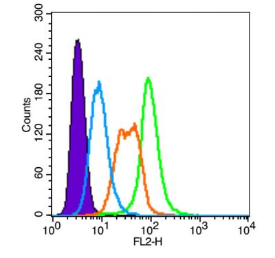 Blank control (blue line): A431(Black). Blank control (blue line): A431(Black).Primary Antibody (green line): Rabbit Anti-phospho-AMPK alpha-1 (Thr172)antibody (bs-4002R) Dilution: 3μg /10^6 cells; Isotype Control Antibody (orange line): Rabbit IgG . Secondary Antibody (white blue line): Goat anti-rabbit IgG-PE(Jackson lab) Dilution: 1μg /test. Protocol The cells were fixed with 4% paraformaldehyde (10 min) , then permeabilized with 90% ice-cold methanol for 20 min on ice. Cells stained with Primary Antibody for 30 min at room temperature. The cells were then incubated in 5%BSA to block non-specific protein-protein interactions followed by the antibody for 15 min at room temperature. The secondary antibody used for 40 min at room temperature. Acquisition of 20,000 events was performed. 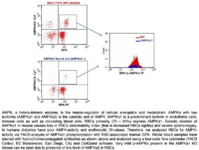 FACS Analysis of Glycophorin A and phospho-AMPK alpha 1/2 (Thr172/183) in Red Blood Cells in WT and AMPK alpha 1 knockout mice using Rabbit Anti-GPA Polyclonal Antibody (bs-2575R-PE) and Rabbit anti-pAMPK alpha1/2 Thr172/183 (bs-4002R-Cy7). Image kindly submitted by Nasrul Hoda, PhD, Georgia Regents University FACS Analysis of Glycophorin A and phospho-AMPK alpha 1/2 (Thr172/183) in Red Blood Cells in WT and AMPK alpha 1 knockout mice using Rabbit Anti-GPA Polyclonal Antibody (bs-2575R-PE) and Rabbit anti-pAMPK alpha1/2 Thr172/183 (bs-4002R-Cy7). Image kindly submitted by Nasrul Hoda, PhD, Georgia Regents University |
我要詢價
*聯(lián)系方式:
(可以是QQ、MSN、電子郵箱、電話等,您的聯(lián)系方式不會被公開)
*內(nèi)容:


