| 中文名稱 | 白細胞介素6受體β/CD130抗體 |
| 別 名 | IL-6R Beta; Interleukin-6 receptor subunit beta; IL-6R-beta; Interleukin-6 signal transducer; Membrane glycoprotein 130; gp130; CDw130; Oncostatin-M receptor subunit alpha; CD_antigen; CD130; GP130 RAPS; gp130 transducer chain; GP130-RAPS; IL6 ST; IL6R-beta; IL6ST.IL6R-beta. |
| 研究領(lǐng)域 | 細胞生物 免疫學(xué) 信號轉(zhuǎn)導(dǎo) 細胞膜受體 |
| 抗體來源 | Rabbit |
| 克隆類型 | Polyclonal |
| 交叉反應(yīng) | Human, Mouse, Rat, |
| 產(chǎn)品應(yīng)用 | WB=1:500-2000 ELISA=1:500-1000 IHC-P=1:100-500 IHC-F=1:100-500 Flow-Cyt=1µg/Test IF=1:100-500 (石蠟切片需做抗原修復(fù)) not yet tested in other applications. optimal dilutions/concentrations should be determined by the end user. |
| 分 子 量 | 99kDa |
| 細胞定位 | 細胞膜 分泌型蛋白 |
| 性 狀 | Liquid |
| 濃 度 | 1mg/ml |
| 免 疫 原 | KLH conjugated synthetic peptide derived from human IL6R beta:651-750/917 |
| 亞 型 | IgG |
| 純化方法 | affinity purified by Protein A |
| 儲 存 液 | 0.01M TBS(pH7.4) with 1% BSA, 0.03% Proclin300 and 50% Glycerol. |
| 保存條件 | Shipped at 4℃. Store at -20 °C for one year. Avoid repeated freeze/thaw cycles. |
| PubMed | PubMed |
| 產(chǎn)品介紹 | CD130 is a signal transducer shared by many cytokines, including interleukin 6 (IL6), ciliary neurotrophic factor (CNTF), leukemia inhibitory factor (LIF), and oncostatin M (OSM). This protein functions as a part of the cytokine receptor complex. The activation of this protein is dependent upon the binding of cytokines to their receptors. vIL6, a protein related to IL6 and encoded by the Kaposi sarcoma-associated herpesvirus, can bypass the interleukin 6 receptor (IL6R) and directly activate this protein. Knockout studies in mice suggested a critical role of the gene encoding this protein in regulating myocyte apoptosis. Alternatively spliced transcript variants encoding distinct isoforms have been described. Function: Signal-transducing molecule. The receptor systems for IL6, LIF, OSM, CNTF, IL11, CTF1 and BSF3 can utilize gp130 for initiating signal transmission. Binds to IL6/IL6R (alpha chain) complex, resulting in the formation of high-affinity IL6 binding sites, and transduces the signal. Does not bind IL6. May have a role in embryonic development. The type I OSM receptor is capable of transducing OSM-specific signaling events. Subunit: Interacts with INPP5D/SHIP1. Forms heterodimers composed of LIPR and IL6ST (type I OSM receptor). Also forms heterodimers composed of OSMR and IL6ST (type II OSM receptor). Homodimer. The homodimer binds two molecules of herpes virus IL6. Component of a hexamer of two molecules each of IL6, IL6R and IL6ST. Interacts with HCK. Subcellular Location: Isoform 1: Cell membrane; Single-pass type I membrane protein. Isoform 2: Secreted. Tissue Specificity: Found in all the tissues and cell lines examined. Expression not restricted to IL6 responsive cells. Post-translational modifications: Phosphorylation of Ser-782 down-regulates cell surface expression. Heavily N-glycosylated. Similarity: Belongs to the type I cytokine receptor family. Type 2 subfamily. Contains 5 fibronectin type-III domains. Contains 1 Ig-like C2-type (immunoglobulin-like) domain. SWISS: P40189 Gene ID: 3572 Database links: Entrez Gene: 3572 Human Entrez Gene: 16195 Mouse Omim: 600694 Human SwissProt: P40189 Human SwissProt: Q00560 Mouse Unigene: 532082 Human Unigene: 706627 Human Unigene: 4364 Mouse Important Note: This product as supplied is intended for research use only, not for use in human, therapeutic or diagnostic applications. |
| 產(chǎn)品圖片 | 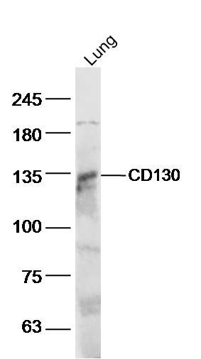 Sample:lung(mouse) Lysate at 40 ug Sample:lung(mouse) Lysate at 40 ugPrimary: Anti- CD130 (bs-1459R) at 1/300 dilution Secondary: IRDye800CW Goat Anti-Rabbit IgG at 1/20000 dilution Predicted band size: 99kD Observed band size: 130 kD 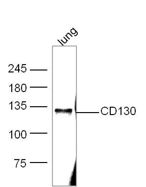 Sample: Lung(Mouse) Lysate at 30 ug Sample: Lung(Mouse) Lysate at 30 ugPrimary: Anti- CD130 (bs-1459R) at 1/300 dilution Secondary: IRDye800CW Goat Anti-Mouse IgG at 1/10000 dilution Predicted band size: 99 kD Observed band size: 130 kD 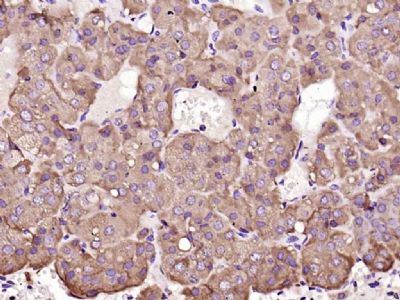 Paraformaldehyde-fixed, paraffin embedded (mouse adrenal gland); Antigen retrieval by boiling in sodium citrate buffer (pH6.0) for 15min; Block endogenous peroxidase by 3% hydrogen peroxide for 20 minutes; Blocking buffer (normal goat serum) at 37°C for 30min; Antibody incubation with (CD130) Polyclonal Antibody, Unconjugated (bs-1459R) at 1:200 overnight at 4°C, followed by operating according to SP Kit(Rabbit) (sp-0023) instructionsand DAB staining. Paraformaldehyde-fixed, paraffin embedded (mouse adrenal gland); Antigen retrieval by boiling in sodium citrate buffer (pH6.0) for 15min; Block endogenous peroxidase by 3% hydrogen peroxide for 20 minutes; Blocking buffer (normal goat serum) at 37°C for 30min; Antibody incubation with (CD130) Polyclonal Antibody, Unconjugated (bs-1459R) at 1:200 overnight at 4°C, followed by operating according to SP Kit(Rabbit) (sp-0023) instructionsand DAB staining.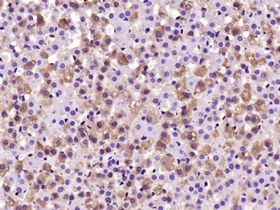 Paraformaldehyde-fixed, paraffin embedded (rat adrenal gland); Antigen retrieval by boiling in sodium citrate buffer (pH6.0) for 15min; Block endogenous peroxidase by 3% hydrogen peroxide for 20 minutes; Blocking buffer (normal goat serum) at 37°C for 30min; Antibody incubation with (CD130) Polyclonal Antibody, Unconjugated (bs-1459R) at 1:200 overnight at 4°C, followed by operating according to SP Kit(Rabbit) (sp-0023) instructionsand DAB staining. Paraformaldehyde-fixed, paraffin embedded (rat adrenal gland); Antigen retrieval by boiling in sodium citrate buffer (pH6.0) for 15min; Block endogenous peroxidase by 3% hydrogen peroxide for 20 minutes; Blocking buffer (normal goat serum) at 37°C for 30min; Antibody incubation with (CD130) Polyclonal Antibody, Unconjugated (bs-1459R) at 1:200 overnight at 4°C, followed by operating according to SP Kit(Rabbit) (sp-0023) instructionsand DAB staining.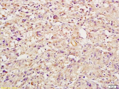 Tissue/cell: Human endometrium carcinoma; 4% Paraformaldehyde-fixed and paraffin-embedded; Tissue/cell: Human endometrium carcinoma; 4% Paraformaldehyde-fixed and paraffin-embedded;Antigen retrieval: citrate buffer ( 0.01M, pH 6.0 ), Boiling bathing for 15min; Block endogenous peroxidase by 3% Hydrogen peroxide for 30min; Blocking buffer (normal goat serum,C-0005) at 37℃ for 20 min; Incubation: Anti-IL-6R Beta/CD130/gp130 Polyclonal Antibody, Unconjugated(bs-1459R) 1:200, overnight at 4°C, followed by conjugation to the secondary antibody(SP-0023) and DAB(C-0010) staining 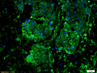 Tissue/cell: human colon carcinoma;4% Paraformaldehyde-fixed and paraffin-embedded; Tissue/cell: human colon carcinoma;4% Paraformaldehyde-fixed and paraffin-embedded;Antigen retrieval: citrate buffer ( 0.01M, pH 6.0 ), Boiling bathing for 15min; Blocking buffer (normal goat serum,C-0005) at 37℃ for 20 min; Incubation: Anti-IL-6R Beta/CD130/gp130 Polyclonal Antibody, Unconjugated(bs-1459R) 1:300, overnight at 4°C; The secondary antibody was Goat Anti-Rabbit IgG, FITC conjugated (bs-0295G-FITC)used at 1:200 dilution for 40 minutes at 37°C. DAPI(5ug/ml,blue,C-0033) was used to stain the cell nuclei 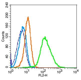 Blank control: Raji(blue). Blank control: Raji(blue).Primary Antibody:Rabbit Anti-CD34 antibody(bs-1459R), Dilution: 1μg in 100 μL 1X PBS containing 0.5% BSA; Isotype Control Antibody: Rabbit IgG(orange) ,used under the same conditions ); Secondary Antibody: Goat anti-rabbit IgG-PE(white blue), Dilution: 1:200 in 1 X PBS containing 0.5% BSA. Protocol The cells were fixed with 2% paraformaldehyde (10 min). Antibody (bs-1459R, 1μg /1x10^6 cells) were incubated for 30 min on the ice, followed by 1 X PBS containing 0.5% BSA + 1 0% goat serum (15 min) to block non-specific protein-protein interactions. Then the Goat Anti-rabbit IgG/PE antibody was added into the blocking buffer mentioned above to react with the primary antibody of bs-1459R at 1/200 dilution for 30 min on ice. Acquisition of 20,000 events was performed. |
我要詢價
*聯(lián)系方式:
(可以是QQ、MSN、電子郵箱、電話等,您的聯(lián)系方式不會被公開)
*內(nèi)容:


